Intracranial Pressure (ICP)

Intracranial pressure (ICP) is the pressure inside the skull and in the brain tissue and cerebrospinal fluid (CSF). If there is a tumor, head injury, brain bleeding, or swelling, the brain can become pressurized like a water balloon, and CSF has nowhere to go. The pressure can crush brain tissue, shift brain structures, contribute to hydrocephalus, cause brain herniation, and restrict blood supply to the brain. In Kiara’s case, elevated ICP caused optic atrophy (blindness due to damage to the optic nerves). ICP is measured in millimeters of mercury (mmHg).

External Ventricular Drain (EVD)

An EVD is a catheter that is inserted by a neurosurgeon into the brain to release cerebrospinal spinal fluid (CSF) and lower intracranial pressure (ICP). It’s basically a tube that drains fluid from inside the brain into a bag next to the bed that can be measured. Drainage can be continuous at a set level, fixed volume per desired time (i.e., every hour), or as needed according to ICP elevations. At the prescribed height, CSF (and/or blood) will drain whenever the interventricular pressure exceeds that set by the height of the collection system. Flow ceases once the pressure equalizes between the CSF compartments in the brain and collection system.

The EVD must be positioned so that that the pressure transducer is in line with the patient’s ear. (Technically, it must point to the Foramen of Monro, which falls at the level of the external auditory meatus of the ear in the supine position and at the mid sagittal line (between the eyebrows) in the lateral position.)  Most nurses “eyeball” it, but some will get a carpenter’s level or laser level to ensure that it’s place accurately. Whenever the patient sits up or lays down, the EVD must be adjusted also, which is a slight — but necessary — hassle.

Hydrocephalus

Hydrocephalus is an abnormal accumulation of cerebrospinal fluid (CSF) within cavities in the brain called ventricles. It typically causes increased pressure inside the skull. Some people may have headaches, double vision, poor balance, urinary incontinence, personality changes, or mental impairment. Other symptoms may include vomiting, sleepiness, seizures, and downward pointing of the eyes.

Hydrocephalus is typically treated by the surgical placement of a shunt system. Before getting a v/p shunt, Kiara had an alternate procedure called a Endoscopic Third Ventriculostomy (ETV) to try to combat the hydrocephalus. It worked well and doctors were fairly confident that there was no increased ICP in her brain, but because of increased vision loss, we opted for a v/p shunt to make sure.

Here is a great video that explains how it works:

Neurosurgeon

A neurosurgeon is a doctor who specializes in the diagnosis and surgical treatment of disorders of the central and peripheral nervous system including congenital anomalies, trauma, tumors, vascular disorders, infections of the brain or spine, stroke, or degenerative diseases of the spine. Less technically speaking, they’re the ones that “go inside” and scoop out what’s not supposed to be there.

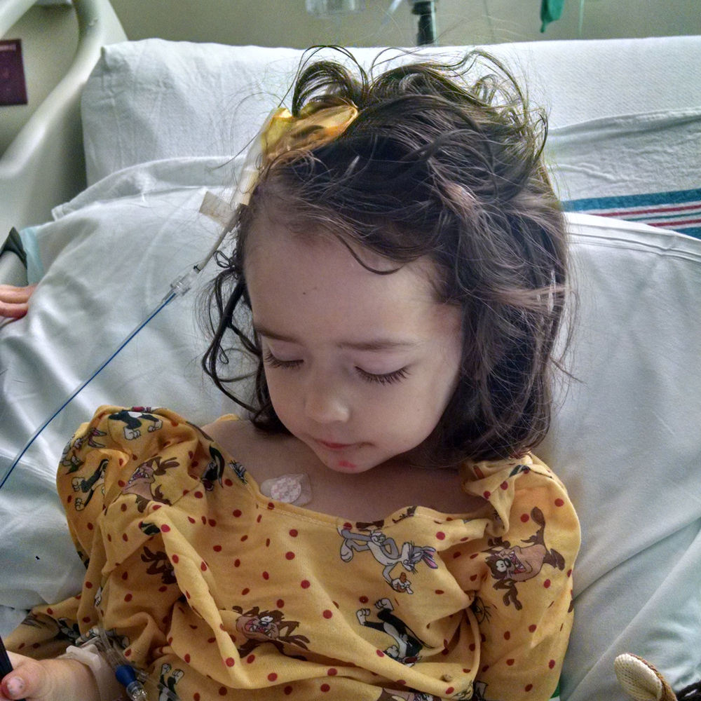
They also have nurse practitioners that do many tasks pre-op and post-op. In Kiara’s case, all of the nurse practitioners are brilliant and beautiful women (seriously, could they be more perfect?). Kiara’s neurosurgeons themselves are unbelievably smart and capable.

Neurosurgeon vs. Neurologist
When a patient has neurological symptoms, often their initial evaluation is with a neurologist, who might obtain imaging to get to the source of the problem. If they discover a structural problem, such as a tumor, they’ll refer the patient to a neurosurgeon. Both doctors treat the same organ, but neurosurgeons operate and neurologists don’t.

Neurologists treat disorders, such as stroke, Alzheimer’s disease, headache, epilepsy, Parkinson’s disease, sleep disorders, multiple sclerosis, pain, tremors, brain, and spinal cord injuries, brain tumors, peripheral nervous disorders and amyotrophic lateral sclerosis. Neurosurgeons treat head and spine trauma and injuries, brain or neck aneurysms or clogged arteries, brain and spinal tumors, and anatomical and structural defects.

Pro Tip: Try to see a neurosurgeon. If you have a clean MRI, then they will refer you to a neurologist or other specialist that can manage your treatment. If their first available appointment isn’t for months, go to the ER and see the neurosurgery nurse practitioner on call. It’s worth it.

MRI

Magnetic resonance imaging (MRI) is a noninvasive medical test that physicians use to diagnose medical conditions. MRI uses a powerful magnetic field, radio frequency pulses and a computer to produce detailed pictures of organs, soft tissues, bone and virtually all other internal body structures. Basically, it’s a big machine that sounds like an automatic Nerf gun and takes pictures of the brain.

Kiara’s scans are done every 3 months or as needed. They take two sets of scans: once without contrast and once with contrast. Neurosurgeons and oncologists use the different types of scans to look for different problems. Tumors show up with contrast. Flair shows up in the scans without contrast. The radiologist is the doctor that first looks over the scans and writes up a report. If there are any questions about the radiology report, the tumor board (consisting of neurosurgeons, oncologist, and others) will review it.

In some operating rooms they will also have an interoperable MRI (IMRI) machine that allows the neurosurgeon to scan the brain during surgery to locate and address any unresolved issues. Unfortunately the IMRI was broken on the day of Kiara’s first major resection, although the neurosurgeons say that it would not have affected the outcome.

Ventriculoperitoneal Shunt

A ventriculoperitoneal (VP) shunt is a medical device that is implanted by a neurosurgeon to treat hydrocephalus and drain excess cerebrospinal fluid (CSF) when the brain ventricles have accumulated too much, potentially causing pressure on the brain. It sits below the skin behind the ear, with one tube gathering CSF from the brain and another tube draining the CSF down into the abdomen, where it is reabsorbed by the body.

Kiara’s vision continued to worsen, so the hypothesis was that there may be increased ICP on her optic nerves. Even though she already had a successful ETV, her neurosurgeon implanted a v/p shunt so that we could be assured that her brain was not a “crock pot of pressure” that could cause her further problems down the line. Although we had been professional shunt-dodgers for a year, we finally consented to the device. Her shunt was manufactured by Certas and it is programmable.

Posterior Fossa Syndrome

I was not prepared for how Kiara would look after her first major tumor resection. Even though her neurosurgeon discussed the likelihood of her having posterior fossa syndrome, it’s difficult to even type the experience. She was trapped in what can only be described as “neuro storms.” Her mouth and tongue would twitch and her eyes jittered separately and in different directions. She was completely unresponsive to me or any of her caregivers like she was in a coma or far, far away. I knew she was in there and probably scared and hurting, and I didn’t leave her side. I thought she may die at any second because her brain and body were just not right. Her head was strongly tilted to the right and her right eye was completely inward. Her lips were puckered. She had tubes everywhere, including an EVD, two IVs, a feeding tube, and a catheter, plus the pulse monitor and blood pressure cuff. Monitors constantly beeped and alarms went off frequently. It was heartbreaking and devastating.

Posterior Fossa Syndrome is a post-operative condition that affects patients that have had surgery on the back side of the base of the skull that contains the brain stem and cerebellum. Symptoms usually appear 1–5 days after surgery. Most symptoms improve in weeks or months, but some symptoms may continue for years following surgery. The symptoms of posterior fossa syndrome include problems with speech and language, motor skills, and mood changes; however, symptoms can be different for each person.

Over the course of a couple of weeks, Kiara slowly regained control of her face, eyes, and body. Speech therapists helped her relearn how to swallow, eat, and speak; physical therapists helped her regain gross motor skills. There was nothing more terrifying than watching her “neuro storms” after her first major resection, and I was so very grateful that she did not have posterior fossa syndrome after her second craniotomy.

Astrocytoma

Juvenile Pilocytic Astrocytoma